Medically reviewed by Dr. Melanie Chin
From the FYidoctors - St. Catharines
Comprehensive Guide: Naming the Parts of the Eye

Comprehensive Guide: Naming the Parts of the Eye
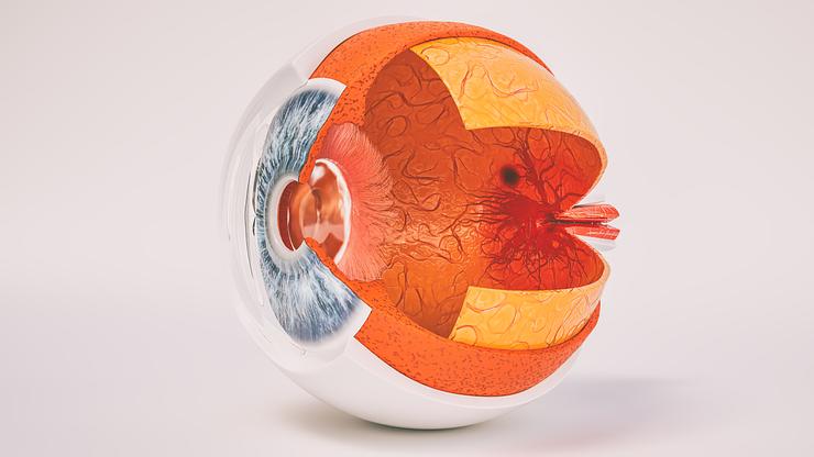
Dive into the human eye's complex structure with this guide, highlighting the essential parts of the eye and their functions. The eye comprises three main layers: the outer layer (sclera and cornea), the middle layer (uvea including the choroid, ciliary body, and iris), and the inner layer (retina). Key components like the cornea and lens work together to focus light, while the photoreceptor cells within the retina convert light into signals sent to the brain. Understanding these parts helps appreciate how vision works and underscores the importance of maintaining eye health through regular exams.
What is the Anatomy of the Eyeball?
The eyeball is composed of three main layers, each playing a crucial role in the visual process:
- The Outer Layer: This layer consists of the sclera and cornea. The sclera is the white, protective outer coat of the eye, while the cornea is the transparent, dome-shaped surface that covers the front of the eye and helps focus incoming light.
- The Middle Layer: Also known as the uvea, this layer includes the choroid, ciliary body, and iris. The choroid contains blood vessels that nourish the eye, the ciliary body controls the shape and focussing ability of the lens and produces aqueous humor (the fluid that keeps the eye pressurized), and the iris is the colored part of the eye that regulates the amount of light entering through the pupil.
- The Inner Layer: This layer is primarily composed of the retina, a light-sensitive tissue that lines the back of the eye. The retina contains photoreceptor cells (rods and cones) that convert light into electrical signals, which are then sent to the brain via the optic nerve.
How Does the Iris Control Light Entering the Eye?
The iris, the colored part of the eye, functions as the eye's aperture, controlling the amount of light that enters the eye. It consists of two types of muscles: the sphincter muscle, which constricts the pupil, and the dilator muscle, which dilates the pupil. When the iris constricts, the pupil becomes smaller, limiting the amount of light entering the eye. Conversely, when the iris dilates, the pupil enlarges, allowing more light to pass through.
This intricate system of pupillary control serves two primary purposes:
- Protecting the eye: In bright light conditions, the iris constricts the pupil to prevent excessive light from entering the eye, which could potentially damage the retina. In low light conditions, the iris dilates the pupil to allow more light to enter, enabling better vision in dimly lit environments.
- Enhancing vision sharpness: By adjusting the pupil size, the iris helps to focus light more precisely onto the retina, improving visual acuity (clarity of vision) and depth of field (how near or far away objects can be while still remaining in focus).
What is the Function of the Retina in Vision?
The retina, the light-sensitive layer at the back of the eye, plays a crucial role in vision by processing light and creating visual images. It contains two types of photoreceptor cells: rods, which are responsible for low-light and peripheral vision, and cones, which handle color vision and fine detail.
When light enters the eye and reaches the retina, the photoreceptor cells convert the light into electrical signals. These signals are then processed by other retinal cells, such as bipolar and ganglion cells, before being transmitted via the optic nerve to the brain, where they are interpreted as visual images.
Within the retina, two specific areas are essential for detailed central vision:
- Macula: This small, central area of the retina is responsible for sharp, clear vision. It contains a high concentration of cone cells, allowing for excellent color perception and visual acuity.
- Fovea: Located at the center of the macula, the fovea is the area of the retina with the highest concentration of cone cells. It enables the most detailed vision, making it crucial for activities such as reading, driving, and recognizing faces.
Understanding the Eye's Protective and Supportive Structures
The eye is equipped with protective and supportive structures that help maintain its health and function. The sclera, the white outer layer of the eye, is a tough, fibrous tissue that provides structure and protection to the delicate inner components. It covers about 80% of the eyeball's surface and is connected to the muscles that control eye movement.
The conjunctiva, a thin, transparent membrane that covers the sclera and lines the inside of the eyelids, plays a crucial role in lubricating and protecting the eye's surface. It contains blood vessels that supply oxygen and nutrients to the eye and produces mucus and tears to keep the eye moist and comfortable. The conjunctiva also helps protect the eye from bacteria, dust, and other foreign particles.
Did You Know? The sclera is thickest at the back of the eye, near the optic nerve, and thinnest at the front, where it meets the cornea. This variation in thickness helps protect the eye from injury while allowing the cornea to remain clear for optimal vision.
Maintaining the health of these protective structures is essential for overall eye health. Regular eye exams can help detect any issues with the sclera or conjunctiva, such as inflammation or infection.
How Do the Lens and Cornea Work Together to Focus?
The lens and cornea are two essential structures that work together to focus light onto the retina, allowing us to see clearly at various distances. The cornea, the clear, dome-shaped surface at the front of the eye, is responsible for refracting or bending light as it enters the eye.
Once light passes through the cornea, it reaches the lens, a transparent, flexible structure that fine-tunes the focusing process. The lens can change its shape, becoming thicker or thinner, to focus on objects at different distances:
- For near vision, the lens becomes more curved, increasing its focusing power.
- For distant vision, the lens flattens, decreasing its focusing power.
The cornea and lens work in harmony to create a clear, focused image on the retina. Each person has a slightly different shaped cornea and lens - structural differences in the shape of the cornea or lens can lead to refractive errors, such as nearsightedness, farsightedness, or astigmatism. Regular comprehensive eye exams with your optometrist can help detect these issues early, allowing for timely treatment to maintain clear vision.
What is the Optic Nerve's Role in Vision?
The optic nerve plays a crucial role in transmitting visual information from the eye to the brain. This bundle of over a million nerve fibers carries electrical impulses generated by the retina's photoreceptor cells (rods and cones) to the visual cortex of the brain for processing.
The optic nerve begins at the optic disc, located at the back of the eye. This small area is where the nerve fibers exit the eye and converge to form the optic nerve – also known as the blind spot. The optic disc lacks photoreceptor cells, which means that any light falling on this spot does not get detected, creating a small gap in the visual field.
However, the brain typically fills in this gap using information from the surrounding areas, so we rarely notice the blind spot in our day-to-day vision. In some cases, damage to the optic nerve, such as from glaucoma, can lead to vision loss or blindness. Regular eye exams are essential for detecting and monitoring the health of the optic nerve to preserve vision.
FAQ
What are the parts of an eyeball?
What are the main parts of the eye and their functions?
What is the black part of the eye called?
What is the outer part of the eye?
What are the three layers and functions of the eyeball?
What causes the white of the eye to turn grey?
What are the three most common visual conditions of the eye and how can each be corrected?

